[vc_row row_type=”0″ row_id=”” blox_height=”” video_fullscreen=”true” blox_image=”” blox_bg_attachment=”false” blox_cover=”true” blox_repeat=”no-repeat” align_center=”” page_title=”” blox_padding_top=”” blox_padding_bottom=”” blox_dark=”false” blox_class=”” blox_bgcolor=”” parallax_speed=”6″ process_count=”3″ video_url=”” video_type=”video/youtube” video_pattern=”true” row_pattern=”” row_color=”” maxslider_image1=”” maxslider_image2=”” maxslider_image3=”” maxslider_image4=”” maxslider_image5=”” maxslider_parallax=”true” maxslider_pattern=”true”][vc_column width=”1/1″][vc_column_text]DEFINITION
Fecal incontinence is the inability to control the methods and times chosen the passage of stool and gas through the anal canal, to discriminate the rectal content and keeping the night control. It ‘a symptom psychologically and socially extremely disabling though not burdened by a high degree of morbidity. It ‘a problem of major social impact with incidence ranging from 0.5 to 5%, although it often remains unrecognized because it is difficult to report because often the patient is ashamed and prefer to hide the problem. The incidence among older people is about 32%, and reaches 56% among older people with neuro-psychiatric disorders.
FISIOPATOLOGIA
La continenza fecale è garantita da un equilibrio tra diversi fattori che sono: la consistenza delle feci, la capacità del serbatorio rettale, la sensibilità rettale e l’efficienza dell’apparato sfinteriale. La mancanza di uno di questi puo’ determinare l’incontinenza.
CAUSES
The main causes of faecal incontinence are diarrhea, fecal impaction, neurological diseases, in case of sphincter apparatus intact. The apparatus anal sphincter alterations lead to fecal incontinence for abnormal operation of the deputies muscular structures to continence, these changes occur as a result of sacrococcigei or pelvic trauma, pelvic surgery and anorectal, seniority, dell’appparato nervous diseases, often after childbirth injuries .
DIAGNOSIS
The first step in the diagnosis of this disease is to report it to your doctor, so you can proceed with the appropriate diagnostic tests. A thorough history and proctologic visit are extremely important to frame the problem in the right way. The symptoms are always collected by means of a simple questionnaire validated at international level that allows measurement of the severity of the problem (Wexner score).[/vc_column_text][vc_column_text]
| Problem | NEVER | 1/Month | 2/Month | 1/Week | 1/Day |
| Gas incontinence | 0 | 1 | 2 | 3 | 4 |
| Liquid feces | 0 | 1 | 2 | 3 | 4 |
| Solid feces | 0 | 1 | 2 | 3 | 4 |
| Soling | 0 | 1 | 2 | 3 | 4 |
| Defecation problems during the day | 0 | 1 | 2 | 3 | 4 |
[/vc_column_text][vc_column_text]Subsequently the diagnostic study must always include the execution of anorectal manometry, a ‘transanal ultrasound and if necessary an electromyography pudendal and a blow-cistus-defecography.
Anorectal manometry records the sphincter tone at rest (which determines the involuntary continence) and voluntary contraction and allows to evaluate the anal sphincter apparatus functions as well as the ampoule rectal sensation. Ultrasound Transanal apparatus evaluates the sphincter integrity and anorectal muscle highlighting any lesions or abnormalities. Electromyography is used to assess the neurological apparatus integrity sphincter and pelvic floor and defecography used to display the function and the anatomical structures of the pelvis during the evacuation.
MEDICAL THERAPY
The first devices have proper education and food hygiene and defecation, so a proper intake of water and fiber in the stool, the use of any medications or supplements constipanti to get a normal consistency of fecal bolus in case of predominantly liquid stools.
Another useful aid consists in the pelvic rehabilitation and biofeeback, the strengthening of the sphincter and pelvic floor muscles techniques.
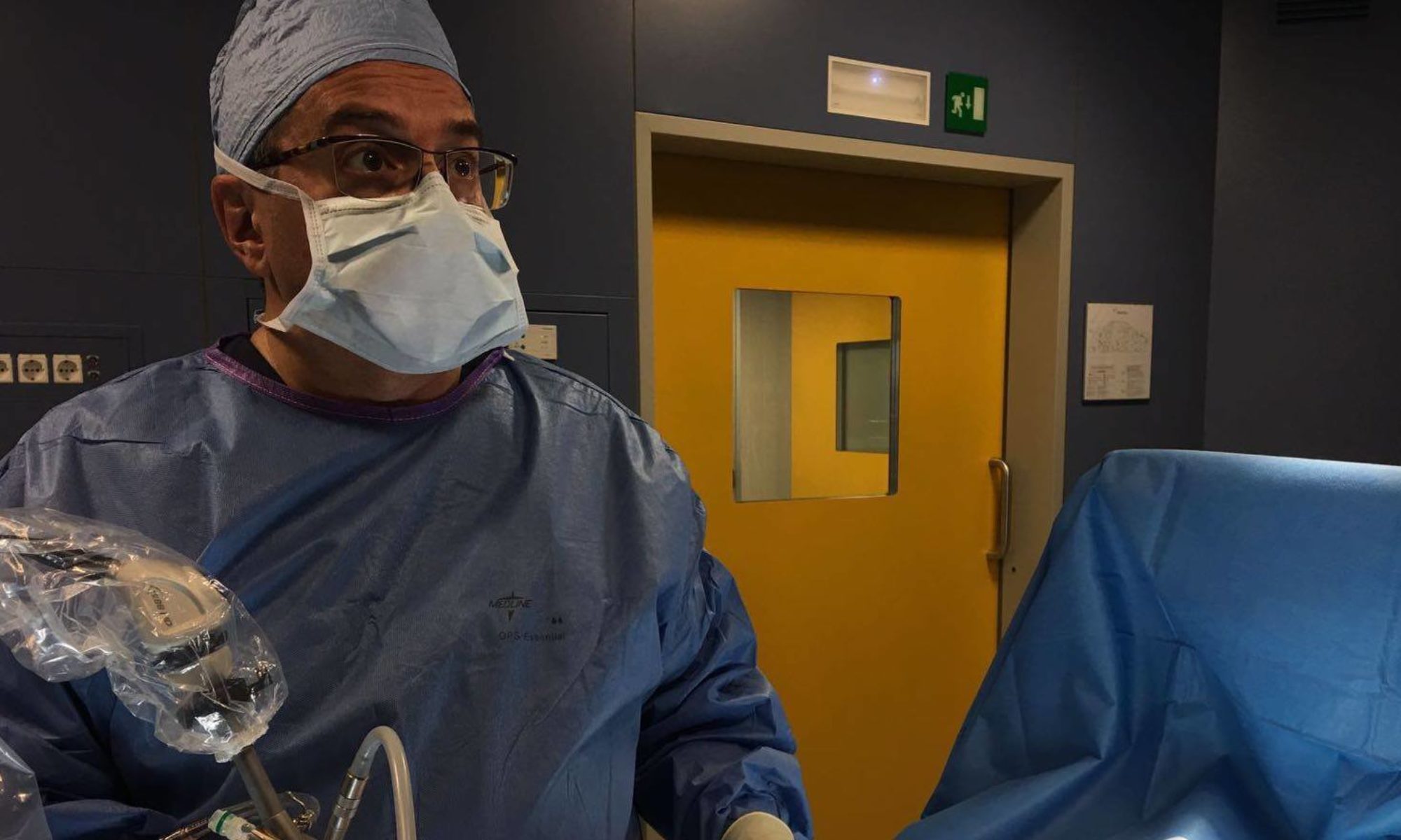
SURGICAL THERAPY
In case of failure of the medical and rehabilitative therapy there are several surgical approaches that are indicated in case of sphincter damage. Each intervention is indicated depending on the type of sphincter damage.
Sphincteroplasty: this operation consists in approaching the separate sphincter stumps from a traumatic injury or surgery.
Graciloplasty: it is to isolate the gracilis muscle (medial aspect of the thigh) which is disposed circumferentially to the anal sphincter. Implantation of artificial anal sphincter.
Sacral neuromodulation: in the plant consists of a stimulator which is to stimulate the sacral nerve through electrical impulses directly to the pelvic muscles and sensitive structures. And ‘an effective therapy to resolve problems with incontinence, pelvic pain and urinary retention. It consists in sending small electrical pulses to a sacral nerve. A lead (that is, a thin wire that ends with 4 small electrodes) is placed next to the appropriate sacral nerve and connected by means of an extension to an implanted stimulator, which sends small electrical pulses to the sacral nerves. The pacing system is implanted under local anesthesia and is composed of:
– Neurostimulator, a small device that is placed in a subcutaneous pocket in the buttock and can ‘be controlled from the outside with a remote control (you can’ turn on or turn off the stimulation or increase and decrease the stimulation).
– Lead placed next to the sacral nerves
The procedure is performed in two stages.
FIRST HALF
For the first time means the percutaneous electrode implant wing. The procedure is performed in the operating room under local anesthesia with radiological monitoring. The electrode is implanted at the level of a sacral foramen (mainly S3) and is brought to the outside through an extension (provisional) which allows a phase of test stimulation in order to assess the effectiveness of treatment.
SECOND HALF
When the test stimulation scored a proven effectiveness and shared on the basis of subjective and objective data of the bladder or bowel diary during the testing phase, we proceed to the connection of the implantable current generator (stimulator) at the top level of the buttock in the subcutaneous with temporary external extension removal.
There are two types of stimulators which differ in the size and battery life. The stimulator duration depends on the stimulation parameters needed and in general is variable between 4 and 10 years. The exhaustion of the battery, the stimulator is replaced with a simple procedure that repeats the step of “the second time”. If unsuccessful after the first time you progress to the electrode removal. These steps have been rare cases of stay of an electrode of the same in the implant site, without listed consequences. In these situations the electrode or the remaining portions may be in place in order to avoid more ‘invasive interventions for removal. The second half expected in the event of therapeutic success the definitive implant stimulator. The definitive implant stimulator is performed under local anesthesia. It removes the temporary external device and connects the electrode to the stimulator that is placed in the subcutaneous; no element of the system will be more visible externally.
In the event of therapeutic failure is removed the neuro modulation system. The same day is on and adjusted the stimulator; also it provides the device’s instructions for use by specialized technical personnel.
When the problem is extremely conditioning and there are no other therapeutic option is used for the packaging of a colostomy.[/vc_column_text][vc_row_inner][vc_column_inner el_class=”” width=”1/3″][ultimate_modal icon_type=”none” modal_title=”Video intervention” modal_contain=”ult-youtube” modal_on=”button” onload_delay=”2″ btn_size=”lg” btn_bg_color=”#325b7b” btn_txt_color=”#ffffff” modal_on_align=”center” btn_text=”Video intervention” txt_color=”#f60f60″ modal_size=”medium” modal_style=”overlay-cornerbottomleft” overlay_bg_color=”#333333″ header_text_color=”#333333″ modal_border_width=”2″ modal_border_color=”#333333″ modal_border_radius=”0″]
[/ultimate_modal][/vc_column_inner][vc_column_inner el_class=”” width=”1/3″][ultimate_modal icon_type=”none” modal_title=”Video intervention” modal_contain=”ult-youtube” modal_on=”button” onload_delay=”2″ btn_size=”lg” btn_bg_color=”#325b7b” btn_txt_color=”#ffffff” modal_on_align=”center” btn_text=”Video intervention” txt_color=”#f60f60″ modal_size=”medium” modal_style=”overlay-cornerbottomleft” overlay_bg_color=”#333333″ header_text_color=”#333333″ modal_border_width=”2″ modal_border_color=”#333333″ modal_border_radius=”0″]
[/ultimate_modal][/vc_column_inner][vc_column_inner el_class=”” width=”1/3″][ultimate_modal icon_type=”none” modal_title=”Video intervention” modal_contain=”ult-youtube” modal_on=”button” onload_delay=”2″ btn_size=”lg” btn_bg_color=”#325b7b” btn_txt_color=”#ffffff” modal_on_align=”center” btn_text=”Video intervention” txt_color=”#f60f60″ modal_size=”medium” modal_style=”overlay-cornerbottomleft” overlay_bg_color=”#333333″ header_text_color=”#333333″ modal_border_width=”2″ modal_border_color=”#333333″ modal_border_radius=”0″]
[/ultimate_modal][/vc_column_inner][/vc_row_inner][vc_empty_space height=”32px”][vc_row_inner el_id=””][vc_column_inner el_class=”” width=”1/3″][ultimate_modal icon_type=”none” modal_title=”Video intervention” modal_contain=”ult-youtube” modal_on=”button” onload_delay=”2″ btn_size=”lg” btn_bg_color=”#325b7b” btn_txt_color=”#ffffff” modal_on_align=”center” btn_text=”Video intervention” txt_color=”#f60f60″ modal_size=”medium” modal_style=”overlay-cornerbottomleft” overlay_bg_color=”#333333″ header_text_color=”#333333″ modal_border_width=”2″ modal_border_color=”#333333″ modal_border_radius=”0″]
[/ultimate_modal][/vc_column_inner][vc_column_inner el_class=”” width=”1/3″][ultimate_modal icon_type=”none” modal_title=”Video intervention” modal_contain=”ult-youtube” modal_on=”button” onload_delay=”2″ btn_size=”lg” btn_bg_color=”#325b7b” btn_txt_color=”#ffffff” modal_on_align=”center” btn_text=”Video intervention” txt_color=”#f60f60″ modal_size=”medium” modal_style=”overlay-cornerbottomleft” overlay_bg_color=”#333333″ header_text_color=”#333333″ modal_border_width=”2″ modal_border_color=”#333333″ modal_border_radius=”0″]
[/ultimate_modal][/vc_column_inner][vc_column_inner el_class=”” width=”1/3″][ultimate_modal icon_type=”none” modal_title=”Get informed consent” modal_contain=”ult-youtube” modal_on=”button” onload_delay=”2″ btn_size=”lg” btn_bg_color=”#325b7b” btn_txt_color=”#ffffff” modal_on_align=”center” btn_text=”Get informed consent” txt_color=”#f60f60″ modal_size=”medium” modal_style=”overlay-cornerbottomleft” overlay_bg_color=”#333333″ header_text_color=”#333333″ modal_border_width=”2″ modal_border_color=”#333333″ modal_border_radius=”0″]
Informed consent neurostimulation
[/ultimate_modal][/vc_column_inner][/vc_row_inner][/vc_column][/vc_row]